A vertically split toenail (onychorrhexis) cannot spontaneously fuse because the nail plate consists of dead keratin. If a split extends back to the cuticle or recurs in the exact same line, the underlying germinal matrix is scarred. While superficial splits can be stabilized with medical fiberglass resin, permanent resolution requires a podiatrist to either microsurgically excise the matrix scar or permanently ablate the offending nail margin. Any vertical split accompanied by a red or dark streak (longitudinal erythronychia) must be professionally evaluated to rule out benign or malignant subungual tumors.
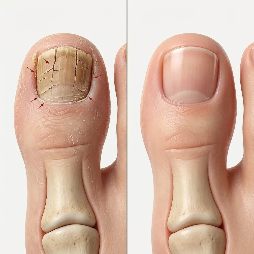
The Anatomy of a Split: Vertical vs. Horizontal Fissuring
Patients frequently confuse two distinct pathological entities when seeking medical guidance for splitting nails:
Horizontal Splitting (Onychoschizia)
Characterized by laminar delamination or peeling of the superficial nail plates at the distal free edge. Caused primarily by extrinsic dehydration, repeated wet-to-dry chemical cycles, harsh solvents, or minor systemic micronutrient insufficiencies (e.g., iron or biotin). It rarely involves the root and readily resolves with topical lipid rehydration and protective lacquers.
Vertical Splitting (Onychorrhexis / Longitudinal Fissure)
A full-thickness parallel crack extending proximally along the longitudinal axis, frequently originating from the germinal matrix beneath the eponychium (cuticle). Because it travels perpendicular to the transverse intercellular tensile force of the nail, it catches aggressively on hosiery, rips into vascular nail bed dermis, and will never heal without structural intervention.
When a vertical split reaches the cuticle, it functions like a zipper: with every step, the mechanical pressure of the shoe and ground reaction force wedges the two nail halves apart, driving the fissure deeper into live tissue.
Etiology: Why the Nail Plate "Zips" Down the Center
The nail plate is continuously manufactured by the germinal matrix, a specialized band of epithelial tissue extending approximately 5 to 8 millimeters beneath the proximal nail fold. Specialized matrix cells (onychocytes) replicate, synthesize hard alpha-keratin, flatten, and push forward along the nail bed.
When you see a persistent central split, one of four biological events has occurred at the matrix level:
- Matrix Laceration or Crush Scarring: Past crush trauma (dropping a heavy weight, severe stubbing, or prior botched partial nail avulsion) lacerates the delicate matrix membrane. In healing, non-nail-producing fibrous scar tissue fills the void. As the surrounding healthy matrix continues extruding nail plate on either side, the scarred gap produces nothing—resulting in a permanent central split.
- Dorsal Pterygium & Lichen Planus: Autoimmune inflammatory disorders can attack the proximal matrix, causing the overlying proximal fold skin to fuse directly down onto the bare nail bed (pterygium unguis), splitting the nail plate into two separate lateral horns.
- Subungual Space-Occupying Mass: A tumor developing within or directly compressive upon the matrix creates a mechanical focal point of failure, indenting or parting the keratin sheet as it emerges.
- Severe Biomechanical Impingement: In patients with hallux limitus, prominent interphalangeal sesamoids, or subungual exostosis (a benign bone spur protruding from the distal phalanx), the bone presses upward from beneath, tenting the nail plate until it fractures longitudinally along its thinnest centerline.
Clinical Differential Matrix: What Does Your Split Signify?
| Presentation Pattern | Suspected Pathology | Diagnostic Hallmark | Primary Podiatric Action |
|---|---|---|---|
| Isolated central split, history of blunt crush trauma | Germinal Matrix Scarring | Split originates at proximal fold; nail is clear and non-painful unless snagged | Matrix Micro-Revision / Phenolization |
| Linear red/pink streak ending in a distal split or notch | Longitudinal Erythronychia (Glomus / SCC) | Point tenderness under cold temperature; solitary digit; expanding red band | Urgent Dermoscopy & Matrix Biopsy |
| Multiple digits, fine longitudinal ridges and splits | Systemic Onychorrhexis / Lichen Planus | Bilateral involvement, "sandpaper" texture (trachyonychia), thinning plates | Dermatologic & Autoimmune Workup |
| Split over a painful dorsal bony bump under nail | Subungual Exostosis | Point pain with tight shoes; firm subcutaneous nodule elevating the center plate | Weight-Bearing X-Ray & Bone Resection |
The Red Flag: Longitudinal Erythronychia & Subungual Tumors
In clinical podiatric dermatology, one specific vertical split pattern triggers an urgent diagnostic workup: Localized Longitudinal Erythronychia (LE).
If your vertical split originates from or runs parallel to a persistent pink, red, or violaceous longitudinal streak that extends from under the cuticle to the free margin, it represents focal matrix thinning caused by an underlying lesion. Key differential conditions include:
- Glomus Tumor: A benign vascular neoplasm originating from the neuromyoarterial glomus bodies in the nail bed. Classically presents with the clinical triad of severe pinpoint tenderness, cold sensitivity (paroxysmal pain in ice water), and a faint red-purple streak with distal splitting.
- Onychopapilloma: The most common benign tumor of the nail bed and distal matrix. Clinically presents with a longitudinal red or brown band, subungual hyperkeratosis (crumbly keratin buildup under the split edge), and a longitudinal fissure.
- Subungual Squamous Cell Carcinoma (SCC) & Bowen's Disease: The most frequent malignant tumor of the nail unit. It is notoriously misdiagnosed as an intractable wart or stubborn fungal split for years before biopsy.
- Amelanotic Subungual Melanoma: A rare variant of acral lentiginous melanoma that lacks dark melanin pigment, presenting instead as an erythematous, vascularized matrix defect with fissuring.
Clinical Rule of Thumb: If a vertical split is accompanied by an unexplained color band, bleeding without trauma, or throbbing pain in cold weather, avoid cosmetic glue patches and book an in-person podiatric dermoscopy evaluation immediately.
Podiatric In-Office Treatment & Surgical Repair Modalities
Depending on whether the split is fresh or chronic, podiatrists deploy a graduated surgical and reconstructive protocol:
1. Medical Fiberglass Splinting & Prosthetic Bridging (Conservative Stabilization)
For distal splits that do not yet involve the root matrix, the goal is mechanical immobilization to prevent the fissure from migrating proximally. Under sterile conditions, the podiatrist debrides the opposing keratin edges, sands the dorsal surface, and bonds a medical-grade fiberglass mesh or biocompatible photopolymer resin across the divide. This acts as an external bridge, allowing the nail to grow forward past the trauma point without snagging.
2. Microsurgical Matrix Scar Revision (Restorative Matrix Reconstruction)
For chronic, through-and-through splits caused by traumatic laceration, surgical reconstruction of the germinal matrix is the gold standard:
- Under local digital block anesthesia, the proximal nail fold is incised laterally and reflected backward to expose the underlying germinal matrix.
- The non-functional linear scar tissue splitting the matrix is meticulously excised under optical magnification with an ultra-fine blade.
- The freshly freed healthy matrix edges are brought into microscopic apposition and sutured with 6-0 or 7-0 rapidly absorbing sutures.
- The nail fold is replaced, and a protective stent or biologic silicone sheet is inserted into the matrix pocket to prevent adhesions during the 9 to 12 months required for the newly unified nail plate to regenerate.
3. Partial Chemical Matrixectomy (The Practical Defect Elimination)
In cases where a split creates a thin lateral fragment (typically 2 to 3 mm on the outer edge) that continually catches, tears into the lateral nail groove, and causes recurring paronychia (bacterial infection), reconstructive suturing is rarely indicated. Instead, the surgeon performs a partial avulsion of the detached sliver and applies 89% chemical phenol to the root matrix for 60 to 90 seconds. This permanently ablates the tiny defective root, leaving a slightly narrower, perfectly uniform nail that never splits again.
At-Home Emergency Stabilization: First Aid Do's and Don'ts
While awaiting your clinical appointment, your primary goal is protecting the raw nail bed from infection and preventing catastrophic tearing on bedsheets or socks:
What TO Do
- Clean with gentle saline or lukewarm soap and water.
- Smooth any sharp, protruding jagged corners with a sanitized fine-grit glass emery board (file in ONE direction only).
- Apply an emergency protective patch using a tiny sterile piece of tea-bag paper saturated with medical-grade cyanoacrylate, placed strictly over intact nail plate.
- Cover with a breathable non-stick bandage before putting on socks.
What NOT To Do
- Never pour industrial hardware superglue into open, weeping, or bloody tissue fissures.
- Never rip, wrench, or yank a hanging half of the nail off with pliers or nail clippers.
- Never pack harsh chemicals like hydrogen peroxide or straight tea tree oil into an open nail fissure.
- Avoid walking barefoot or wearing tight-fitting slip-on shoes that apply transverse torque to the toe.
Stop Living With a Painful, Snagging Split Toenail
A chronic longitudinal split will not fix itself. Our network of board-certified foot and ankle surgeons provide gentle digital numbing, immediate pain relief, and permanent reconstructive solutions covered by most insurances or flat-rate transparent cash pricing.
Frequently Asked Questions
Q Can a toenail that is split down the middle heal back together on its own?
Q Why does my toenail keep splitting in the exact same vertical line every time it grows?
Q What is longitudinal erythronychia and when does a split indicate a tumor?
Q Can I use superglue or nail glue to fix a split toenail at home?
Q What does a podiatrist do to permanently repair a vertically split toenail?
Related Clinical Analysis
Onycholysis Overview
Clinical causes of nail plate separation from the distal nail bed.
Subungual Hematoma
Trauma-induced bleeding beneath the nail plate leading to structural fissures.
Melanonychia & Pigment Bands
Longitudinal nail pigmentation differentials and subungual melanoma screening.
Top Podiatrists for Nail Repair
Consult with board-certified podiatric surgeons for permanent matrix repair.