Toenail Discoloration From Medications
Find a Podiatrist Near You
Get same-day appointments with verified podiatrists. Insurance accepted.
Toenail discoloration from medications is one of the most consistently misdiagnosed nail presentations in clinical practice — because the nail changes that drugs produce look remarkably similar to the changes caused by fungal infection, trauma, or systemic disease. A patient on chemotherapy who notices dark bands developing across their nails, or someone taking doxycycline who sees their toenails yellowing, will almost always assume something has gone wrong with their nails. Very rarely do they connect the change to a medication they may have been taking for weeks or months.
This matters because toenail discoloration from medications typically does not require treatment of the nail itself — it requires understanding the mechanism, knowing whether it is reversible, and knowing when it genuinely warrants physician review versus when it should simply be monitored as a known and expected side effect. And it matters because prescribing or self-treating with antifungal medication for what is actually a drug-induced nail change wastes time and can delay identifying the actual cause.
This guide explains eight categories of medications that cause toenail discoloration, the specific mechanisms behind each color change, how to distinguish drug-induced changes from other causes, and what appropriate management looks like.

How Medications Cause Toenail Discoloration
Toenail discoloration from medications can occur through several distinct biological mechanisms — and the mechanism determines the color change, the pattern, and whether it reverses when the medication stops.
Drug pigment deposition:
Some medications or their metabolites are directly deposited into the keratin of the nail plate as it grows. Since the nail plate is built from matrix cells that incorporate whatever is present in the surrounding tissue, drugs that accumulate in tissues can become physically embedded in the nail plate structure. This produces pigmentation that is within the nail plate itself — the color is in the structure, not on the surface.
Melanocyte activation:
Certain medications activate nail matrix melanocytes — the pigment-producing cells — stimulating them to deposit more melanin than normal into the growing nail plate. This produces longitudinal or diffuse dark pigmentation of the nail.
Photosensitivity-induced nail bed damage:
Some medications concentrate in peripheral tissues including the nail bed and make those tissues abnormally sensitive to UV light. Sun exposure triggers a phototoxic reaction in the sensitized nail bed, producing inflammatory changes that alter the nail’s color and, in some cases, its attachment to the nail bed.
Vascular effects on nail bed color:
Some medications alter peripheral circulation or red blood cell characteristics, changing how the nail bed appears through the translucent nail plate.
Nail matrix toxic effects:
Cytotoxic medications — particularly chemotherapy agents — can disrupt nail matrix cell production in ways that alter the structural composition of the nail plate, affecting how it reflects and transmits light.
Understanding which mechanism a specific medication uses helps predict what the nail changes will look like and whether they will resolve when the drug is stopped.
8 Medication Categories Causing Toenail Discoloration
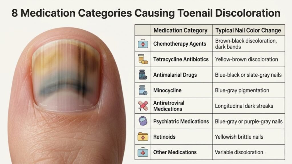
1. Chemotherapy Agents — The Most Extensive Nail Effects
Chemotherapy drugs produce the widest range and most dramatic toenail discoloration from medications, because cytotoxic agents affect all rapidly dividing cells — and the nail matrix is one of the most rapidly dividing tissues in the body.
Mechanisms:
- Direct matrix toxicity disrupting normal keratin cell production
- Melanocyte activation from matrix inflammatory response
- Drug pigment incorporation into the growing nail plate
Specific nail changes by drug class:
Taxanes (docetaxel, paclitaxel):
One of the most commonly documented causes of severe nail toxicity from chemotherapy. Taxanes produce multiple nail changes simultaneously: diffuse brownish-black nail discoloration, Beau’s lines (horizontal grooves from matrix disruption), onycholysis (nail lifting), and in severe cases, subungual hematomas without trauma. The nail changes typically appear after several treatment cycles and can affect all nails simultaneously.
Hydroxyurea:
Produces longitudinal melanonychia — dark vertical streaks running from nail base to tip across multiple nails. The mechanism involves hydroxyurea-induced melanocyte activation in the nail matrix. The streaks may be brown or dark gray and affect all nails relatively symmetrically.
Bleomycin:
Produces brown or dark banding across multiple nails — horizontal bands corresponding to treatment cycles, similar to Beau’s lines but pigmented. Also associated with nail thickening and onycholysis.
Doxorubicin (Adriamycin) and anthracyclines:
Diffuse nail darkening and nail band formation.
EGFR inhibitors (erlotinib, cetuximab, gefitinib):
A newer class of targeted cancer therapy producing characteristic paronychia (nail fold inflammation) alongside nail changes including discoloration, ingrown nail tendencies, and nail fragility.
Clinical significance:
Chemotherapy-induced toenail discoloration from medications is expected and documented — it does not indicate additional disease. However, severe nail changes including significant onycholysis or painful paronychia may require nail care intervention and discussion with the oncology team about dose modification.
2. Tetracycline Antibiotics — Photosensitivity Mechanism
Tetracycline antibiotics — particularly doxycycline — are one of the most commonly encountered causes of toenail discoloration from medications in otherwise healthy patients taking antibiotics for acne, Lyme disease, or infections.
Mechanism — photosensitization:
Tetracyclines concentrate in peripheral tissues including the nail bed. When UV light (from sun exposure) reaches these tissues, it triggers a phototoxic reaction — the drug absorbs light energy and generates reactive oxygen species that damage the nail bed tissue and disrupt nail plate attachment. This produces both nail discoloration and nail separation (photo-onycholysis).
What tetracycline nail changes look like:
- Yellow or brownish-yellow nail discoloration
- Photo-onycholysis — nail lifting at the nail tip, most pronounced on nails most exposed to sunlight
- Bilateral and symmetric pattern (both feet, same nails affected)
- The changes are typically worse in summer or after significant sun exposure
- All nails on sun-exposed extremities may be affected
Clinical significance:
Tetracycline-induced photo-onycholysis can be confused with fungal infection. The temporal correlation with doxycycline use, the symmetric bilateral pattern, and the improvement with sun protection distinguish it from fungal causes.
Management:
Sun protection — covering feet and keeping them out of direct sunlight while on tetracycline antibiotics can prevent or limit photo-onycholysis. If the nail separation is significant, discussing alternative antibiotics with the prescribing physician is appropriate.
3. Antimalarial Drugs — Blue-Black Pigmentation
Hydroxychloroquine and chloroquine — antimalarial medications also widely used for rheumatoid arthritis and lupus — produce a distinctive blue-black or gray-black nail pigmentation as a recognized long-term side effect.
Mechanism:
Antimalarial drugs form complexes with melanin and accumulate in melanin-containing tissues over time. Nail matrix melanocytes produce melanin that incorporates drug-melanin complexes, which are then deposited into the growing nail plate. The resulting pigmentation is blue-black or gray-brown — distinct from the yellower pigmentation of fungal infection.
What antimalarial nail changes look like:
- Blue-black or slate-gray diffuse nail discoloration
- May also affect the skin (blue-gray skin discoloration — chrysoderma) and the retina
- Typically develops after months to years of treatment, not with short-term use
- Affects multiple nails symmetrically
- The discoloration may also produce horizontal banding on the nail corresponding to drug concentration patterns
Clinical significance:
Patients on long-term hydroxychloroquine for lupus or rheumatoid arthritis who notice nail darkening should report it to their prescribing physician. The degree of nail pigmentation can correlate with tissue drug accumulation levels.
4. Minocycline — The Distinctive Blue-Gray Change
Minocycline, another tetracycline-class antibiotic used for acne and certain infections, produces nail discoloration through a different mechanism from the photo-onycholysis of doxycycline.
Mechanism:
Minocycline forms stable complexes with iron and melanin in tissues. Over time, these minocycline-metal complexes accumulate in multiple tissues — the skin (producing the characteristic blue-gray or slate-gray skin discoloration of long-term minocycline use), the gums, and the nails.
What minocycline nail changes look like:
- Blue, blue-gray, or slate discoloration of the nail plate
- Affects multiple nails
- Associated with similar blue-gray discoloration in skin (particularly sun-exposed areas like the shins and face) and gum tissue
- Develops after extended periods of use — typically months to years
- The discoloration may persist for months to years after the medication is discontinued because the drug-metal complexes are slow to clear from tissues
Clinical significance:
The combination of blue-gray nail discoloration + skin discoloration + gum discoloration in a patient on long-term minocycline is pathognomonic (uniquely characteristic). The drug should be discussed with the prescribing physician.
5. Antiretroviral Medications — Melanonychia in HIV Treatment
Zidovudine (AZT) — an antiretroviral medication used in HIV treatment — is one of the most documented causes of toenail discoloration from medications in the form of drug-induced melanonychia (dark longitudinal streaks).
Mechanism:
Zidovudine activates nail matrix melanocytes, stimulating them to produce and deposit melanin into the growing nail plate. This produces longitudinal melanonychia — dark vertical streaks — affecting multiple nails.
What AZT nail changes look like:
- Longitudinal brown or dark bands running from nail base to tip
- Affects multiple nails simultaneously — often all nails
- May also produce diffuse nail darkening rather than discrete bands
- The pattern is symmetric — both hands and feet affected similarly
Important clinical consideration:
HIV-positive patients have elevated baseline rates of benign melanonychia even without antiretroviral medication. They also have elevated risk of subungual melanoma compared to the general population. New dark streaks in HIV-positive patients should be evaluated professionally rather than assumed to be medication-related — particularly if the pattern is atypical or involves single-nail involvement with concerning features (widening, Hutchinson’s sign).
Other antiretroviral medications:
Other drugs in the HIV treatment regimen can produce nail effects including discoloration, nail fragility, and onycholysis as class effects.
6. Psychiatric Medications — Phenothiazine-Class Pigmentation
Phenothiazine antipsychotics — particularly chlorpromazine — produce a distinctive pigmentation pattern as a long-term side effect that affects skin, eyes, and nails.
Mechanism:
Phenothiazines and their metabolites accumulate in melanin-containing tissues and in sun-exposed tissues. They produce a blue-gray to purple discoloration in the skin (particularly sun-exposed areas), the cornea and lens, and the nails.
What phenothiazine nail changes look like:
- Blue-gray or purple-gray nail discoloration
- Occurs alongside similar skin pigmentation in sun-exposed areas
- Long-term treatment association
- May be difficult to distinguish from minocycline pigmentation without medication history
Clinical significance:
This represents a long-term accumulation effect rather than a dose-related effect. The pigmentation may be very slow to fade even after the medication is discontinued.
7. Retinoids — Nail Matrix Effects
Retinoids — including isotretinoin (for severe acne) and acitretin (for psoriasis) — produce nail changes through their effects on keratin production and nail matrix cell behavior.
Mechanism:
Retinoids regulate keratin gene expression. In the nail matrix, this produces changes in the structural organization of keratin, altering the nail plate’s optical properties. They also produce paronychia-like inflammatory changes in some patients.
What retinoid nail changes look like:
- Nails may appear more yellow and less lustrous
- Nail plate may become thinner and more fragile
- Paronychia — inflammation of the nail fold — is a recognized retinoid side effect
- Brittle nails that break and peel more readily
- These changes are dose-dependent and typically improve when the retinoid is discontinued or reduced
8. Other Medications With Documented Nail Discoloration Effects
Several additional medication classes produce toenail discoloration from medications through various mechanisms:
Psoralens (used in PUVA therapy):
Psoralens are photosensitizing compounds used alongside UV light for skin conditions including psoriasis. They produce nail changes through photosensitization — yellow nail discoloration and increased nail brittleness, similar to but more pronounced than tetracycline photosensitization.
Some NSAIDs (non-steroidal anti-inflammatory drugs):
Certain NSAIDs in high doses or long-term use have been associated with photo-onycholysis and nail discoloration, though this is less common than with tetracyclines.
Some cardiovascular medications:
Beta-blockers and certain other cardiovascular drugs have occasional case reports of nail discoloration, though the evidence base is limited compared to the medications above.
Silver-containing medications:
Historically, silver-based medications accumulated in tissues producing argyria — blue-gray pigmentation of skin and nails. This is now rare with modern medications but may occur with colloidal silver products purchased as supplements.
Distinguishing Toenail Discoloration From Medications vs Other Causes
| Feature | Drug-Induced Discoloration | Fungal Infection | Trauma-Induced |
|---|---|---|---|
| Nails affected | Multiple, often all | Often starts in one | Typically one or two |
| Pattern | Symmetric, bilateral | Asymmetric spread | Specific nail |
| Color | Varies by drug (dark bands, blue-gray, yellow) | Yellow, brown, white | Dark red, purple, brown |
| Subungual debris | Absent | Present — chalky debris | Absent |
| Nail crumbling | Absent (unless severe chemo toxicity) | Present | Absent |
| Nail lifting | Possible (photo-onycholysis) | Present in advanced infection | Possible |
| Medication history | Present | Not required | Not required |
| Timing | Corresponds with medication start | Progressive over months | After specific injury |
| Lab test (KOH/culture) | Negative | Positive | Negative |
What to Do When You Notice Toenail Discoloration From Medications

Step 1: Connect the timing
When did you first notice the nail change? How long have you been on the suspected medication? Toenails grow approximately 1.5 mm per month — a color change appearing 6 weeks after starting a medication is plausible for a drug-induced effect.
Step 2: Check which nails are affected
Multiple nails affected symmetrically strongly suggests a systemic cause — drug-induced or systemic disease — rather than fungal infection or trauma, which typically start asymmetrically.
Step 3: Describe the color specifically
Yellow nails → more consistent with tetracyclines, retinoids, or fungal infection
Brown/dark bands → more consistent with chemotherapy, antiretrovirals, hydroxyurea
Blue-gray → more consistent with minocycline, antimalarials, phenothiazines
Green → bacterial colonization (Pseudomonas) — separate from drug effects
Step 4: Do not stop medication independently
Never discontinue a prescribed medication because of nail discoloration without discussing it with your prescribing physician. The medication may be essential for your health, and there may be alternatives that produce fewer nail effects, or the nail change may be acceptable and manageable without medication change.
Step 5: Report to your prescribing physician
Document the nail changes with photographs and bring them to your next appointment. This is exactly the type of side effect that your prescribing physician needs to know about.
Management of Toenail Discoloration From Medications
For most cases of drug-induced nail discoloration:
No specific nail treatment is needed. The discoloration is within the nail plate and will grow out as new nail replaces the affected portion over 9 to 18 months. The key is identifying the medication as the cause so that inappropriate treatment (antifungals) is avoided.
For photo-onycholysis from tetracyclines or psoralens:
Strict sun protection — keeping feet covered and out of direct sunlight — can prevent further progression. If already established, reducing UV exposure allows new nail to grow normally from the matrix.
For severe chemotherapy-related nail toxicity:
Nail cooling during chemotherapy infusions (cryotherapy) is an evidence-based preventive measure that reduces blood flow to the nail bed during drug administration, limiting drug concentration at the nail matrix. Nail care with regular gentle trimming, moisturizing, and protection from secondary infection is important throughout treatment.
For minocycline or antimalarial blue-gray pigmentation:
These changes may be slow to resolve — months to years after medication discontinuation — because the drug-metal complexes clear slowly from tissues. Discussing alternative medications with the prescribing physician is the most effective management if the pigmentation is cosmetically or clinically significant.
Frequently Asked Questions About Toenail Discoloration From Medications
Can medications really change toenail color?
Yes. Multiple medication classes produce documented, recognized nail discoloration as side effects. Chemotherapy, tetracycline antibiotics, antimalarials, antiretrovirals, minocycline, retinoids, and phenothiazines are among the most commonly implicated.
How do I know if my nail discoloration is from medication or fungal infection?
The key distinguishing features are: multiple nails affected symmetrically (suggests medication/systemic cause), timing correlated with medication start, absence of subungual debris beneath the nail (absent in drug-induced, present in fungal), and negative laboratory fungal testing. Professional evaluation confirms the cause.
Will stopping the medication fix my toenail discoloration?
For most drug-induced nail discoloration, stopping the medication allows new nail to grow normally from the matrix — the discolored nail then grows out over 9 to 18 months. Some changes (minocycline blue-gray, antimalarial pigmentation) may be slow to resolve even after stopping. Never stop a prescribed medication without physician guidance.
Should I be treated for fungal infection if my nails are discolored from medication?
No — if the discoloration is confirmed to be drug-induced, antifungal treatment is not appropriate and will not help. The cause needs to match the treatment.
Are chemotherapy nail changes dangerous?
Chemotherapy-related nail discoloration itself is not dangerous. However, severe nail toxicity from chemotherapy — significant onycholysis, painful paronychia, nail loss — may require nail protective care and discussion with your oncology team about dose adjustments.
How long does medication-induced nail discoloration last?
Nail discoloration from medications typically resolves as new nail grows from the matrix over 9 to 18 months after the medication is stopped. Minocycline and antimalarial pigmentation may persist longer due to slow tissue clearance of the drug.
Summary
Toenail discoloration from medications affects patients across many medical specialties — oncology, dermatology, rheumatology, infectious disease, and psychiatry. The eight most important medication categories are chemotherapy agents, tetracycline antibiotics, antimalarials, minocycline, antiretrovirals, retinoids, phenothiazines, and psoralens — each producing nail discoloration through distinct mechanisms that determine the color, pattern, and reversibility of the change.
The clinical challenge is distinguishing toenail discoloration from medications from fungal infection, which these changes can closely resemble. The key differentiating features are the symmetric multi-nail pattern, temporal correlation with medication use, absence of subungual debris, and negative fungal laboratory testing.
Management centers on identifying the medication as the cause, avoiding unnecessary antifungal treatment, monitoring nail changes, and reporting them to the prescribing physician. In most cases, the discoloration resolves gradually as new nail grows out after the medication is stopped or changed — though some drug-pigment deposits clear more slowly than others.
Struggling to get an appointment?
We help patients in your area bypass clinic waitlists by instantly finding verified podiatry doctors who accept their insurance.